Thyroid Ultrasound: How Imaging Nodules Reveals Cancer Risk

Thyroid ultrasound is the most common test doctors use to check for nodules in the thyroid gland-and it’s not just about finding lumps. It’s about figuring out which ones might be cancerous and which ones are harmless. Unlike physical exams, where a doctor feels your neck, ultrasound can spot nodules as small as 2 millimeters. Studies show that up to 68% of people have thyroid nodules when scanned, but only about 5-10% of those turn out to be cancer. The real job of ultrasound isn’t to diagnose cancer-it’s to tell you which nodules need a closer look.
How Thyroid Ultrasound Works
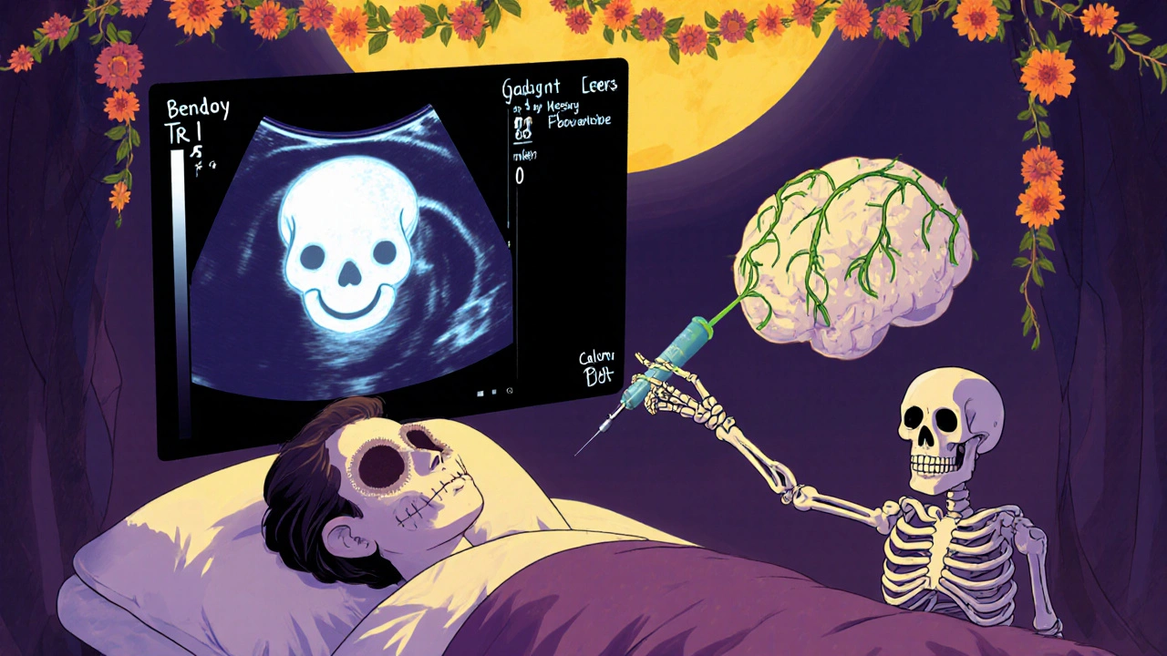
Thyroid ultrasound uses high-pitched sound waves, not radiation, to build a real-time image of your thyroid. A handheld device called a transducer is moved over your neck, sending out waves that bounce off tissues and create a picture on a screen. The machine operates at frequencies between 7.5 and 18 MHz-higher frequencies give sharper images of shallow structures like the thyroid.
Modern systems also include Doppler technology, which shows blood flow inside nodules. Cancerous nodules often have abnormal blood vessels that run through the center, not just around the edges. This is one of the red flags radiologists watch for. The whole process takes 15 to 30 minutes, costs between $200 and $500 in the U.S., and requires no prep or recovery time.
The Five Key Features That Raise Red Flags
Not all nodules are created equal. Radiologists look at five specific traits to judge risk:
- Composition: Is the nodule mostly fluid (cystic), full of tiny holes (spongiform), or solid? Solid nodules carry more risk than cysts.
- Echogenicity: How bright or dark does it look compared to the surrounding thyroid tissue? Markedly hypoechoic (very dark) nodules are more likely to be cancerous.
- Shape: A nodule that’s taller than it is wide (like a vertical oval) is a major warning sign. Normal nodules are wider than they are tall.
- Margin: Smooth edges are good. Jagged, blurry, or spreading beyond the thyroid border are bad.
- Punctate echogenic foci: Tiny white dots inside the nodule-called microcalcifications-are one of the strongest predictors of cancer.
These five features are the foundation of TI-RADS, the system doctors use to rate cancer risk. Each feature gets 0 to 3 points based on how suspicious it looks. Add them up, and you get a score that tells you your risk level.
Understanding TI-RADS Risk Levels
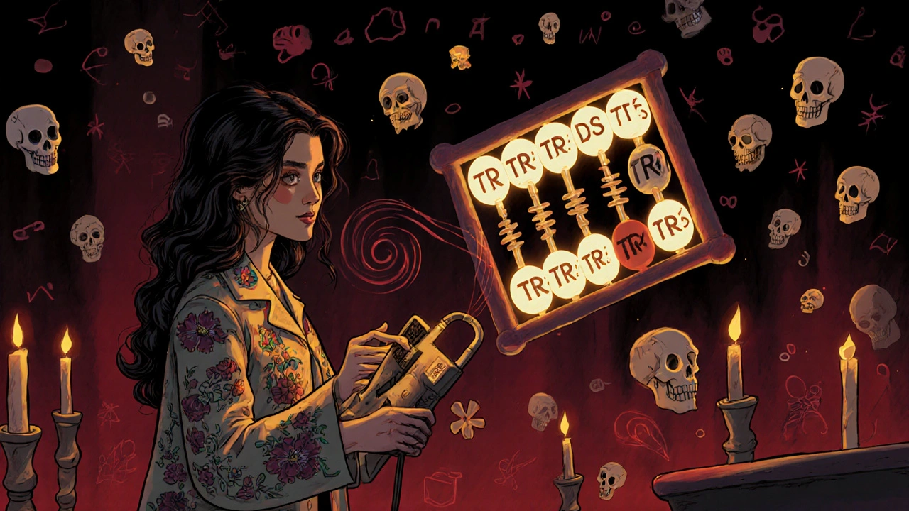
The Thyroid Imaging Reporting and Data System (TI-RADS) was created by the American College of Radiology to bring consistency to how doctors interpret ultrasounds. Here’s what the scores mean:
- TR1 (0 points): 0.3% chance of cancer. No follow-up needed.
- TR2 (2 points): 1.5% chance. Routine monitoring, no biopsy.
- TR3 (3 points): 4.8% chance. Biopsy if nodule is over 1 cm.
- TR4 (4-6 points): 9.1% chance. Biopsy recommended for nodules 1 cm or larger.
- TR5 (7+ points): 35% chance. High suspicion. Biopsy is almost always needed.
For example, a solid, very dark nodule with microcalcifications and a taller-than-wide shape could easily hit 7 points-putting it in TR5. That’s a strong signal to move forward with a biopsy. But a small, fluid-filled nodule with smooth edges and no calcifications? That’s TR1. You’ll likely never hear from it again.

Why Ultrasound Beats Other Tests
You might wonder: Why not just get a CT scan or MRI? Those can show a nodule, but they can’t tell you if it’s dangerous. They don’t show microcalcifications, shape, or blood flow patterns. Nuclear scans (like radioactive iodine uptake tests) can say if a nodule is "hot" (overactive) or "cold" (underactive), but only cold nodules have any real cancer risk-and even then, only about 15% of them are malignant. And those scans expose you to radiation.
Ultrasound is the only test that gives you the full picture without radiation. It’s also the only way to guide a needle during a biopsy. When doctors use ultrasound to steer the needle into a nodule, they get good tissue samples 95% of the time. Without it, up to 25% of biopsies come back useless because they missed the nodule entirely.
When Biopsy Is-and Isn’t-Needed
Not every nodule needs a biopsy. Size matters, but so does risk level.
- Nodules under 5 mm: Even if they look suspicious, skip the biopsy. They’re almost never dangerous.
- Nodules 1 cm or larger with TR4 or TR5 features: Biopsy is recommended.
- Nodules 2.5 cm or larger with TR3 features: Biopsy is advised. Studies show thyroid cancers don’t affect survival until they reach this size.
Even after a biopsy, you might still need follow-up. About 15-30% of biopsies come back "indeterminate"-meaning the cells look unusual but not clearly cancerous. In these cases, molecular testing (like Afirma or ThyroSeq) can help. These tests look at gene mutations and can rule out cancer in many cases, avoiding unnecessary surgery. But even if the test says benign, doctors still recommend yearly ultrasounds to watch for changes.
What Happens After a Cancer Diagnosis?
Most thyroid cancers are papillary, slow-growing, and highly treatable. For small cancers under 1 cm, many doctors now recommend active surveillance instead of immediate surgery. A 10-year survival rate of over 99% has been seen in patients who choose monitoring over removal. Surgery is still an option, but it’s no longer the automatic first step.
Ultrasound plays a role here too. After surgery, it’s used to check for lymph node spread or recurrence. It’s also the go-to tool for tracking nodule growth over time-even if the biopsy was benign.

Challenges and Limitations
Ultrasound is powerful, but it’s not perfect. One big problem? Different doctors see things differently. Studies show only moderate agreement between radiologists when judging margins or echogenicity. This is why training matters. To be consistent, radiologists need to read 200 to 300 scans under supervision.
Another issue: many clinics don’t check lymph nodes properly. About 35% of community ultrasounds miss this step. But cancer can spread to lymph nodes in the neck before it grows large. A good ultrasound should always include a full neck survey.
Equipment matters too. A machine with a 10 MHz linear transducer, Doppler, and compound imaging is the minimum standard. Cheap or outdated machines can miss key details.
The Future: AI and Personalized Risk
Artificial intelligence is starting to change thyroid ultrasound. A 2023 study in Nature Scientific Reports tested a deep learning model that analyzed nodule shape, texture, and blood flow. It reached 94.2% accuracy-better than most human radiologists. The model caught patterns even experts missed.
Next up? Combining ultrasound features with genetic markers. Imagine a system that looks at your nodule’s shape, calcifications, and DNA profile all at once to give you a personalized cancer risk score. Experts predict this will cut unnecessary biopsies by 30% in the next five years.
For now, ultrasound remains the gold standard. No other test matches its safety, speed, detail, or ability to guide treatment. As technology improves, it will only get better.
Frequently Asked Questions
Is thyroid ultrasound safe?
Yes. Thyroid ultrasound uses sound waves, not radiation, so it’s completely safe. It’s safe for pregnant women, children, and people who need repeated scans. There are no known side effects.
Can thyroid ultrasound diagnose cancer?
No. Ultrasound can only estimate cancer risk based on nodule features. Only a biopsy can confirm cancer. Ultrasound tells you which nodules are worth biopsying-but it doesn’t give a final diagnosis.
Do all thyroid nodules need treatment?
No. Most thyroid nodules are benign and don’t need any treatment. Many are simply watched with yearly ultrasounds. Only nodules that are suspicious on ultrasound, large, or causing symptoms require biopsy or surgery.
How often should I get a thyroid ultrasound?
If your nodule is low-risk (TR1 or TR2), you typically don’t need another scan unless it grows or you develop symptoms. For TR3 nodules, follow-up ultrasound is usually recommended in 12 to 24 months. Higher-risk nodules (TR4 or TR5) may need scans every 6 months before biopsy.
What if my biopsy is inconclusive?
If the biopsy result is indeterminate, molecular testing can help clarify the risk. If that still doesn’t give a clear answer, doctors often recommend close ultrasound monitoring every 6 months for 2-3 years. Surgery may be considered if the nodule grows or new suspicious features appear.


I got a thyroid nodule last year and they told me it was TR3... I cried for three days. Then I Googled 'thyroid cancer survival rates' and found a guy who said he lived on kombucha and yoga for 12 years after diagnosis. Now I drink it every morning. 🥤✨
You know what's wild? The fact that we're using sound waves to detect microscopic white dots inside a gland the size of a butterfly's wing. And yet, we still can't reliably diagnose depression with a blood test. Medicine is equal parts miracle and magic. Also, if your radiologist doesn't check lymph nodes, fire them. That's like hiring a chef who forgets to taste the soup.
THIS IS A GOVERNMENT COVER-UP. Ultrasound doesn't detect cancer - it detects the 5G signals they implant in your neck during flu shots. The 'microcalcifications'? That's nanotech tracking devices. They're using thyroid scans to map who's 'high risk' for the next vaccine mandate. I've seen the documents. They're hiding it in plain sight.
If you're over 40 and haven't had a thyroid scan, you're just gambling with your life. 😔
Why are we letting foreign tech companies control the AI models that read our scans? We need American-made ultrasound software. Made in the USA. Built by patriots. Not some Silicon Valley startup funded by Chinese venture capital.
In India, we don't even have access to this level of tech in most towns. My cousin got a nodule detected because his aunt saw a bump while combing his hair. No ultrasound. No TI-RADS. Just prayers and turmeric milk. Still alive at 78. 🙏🌿
I read this whole thing and now I'm convinced I have TR5. I've been googling 'thyroid cancer symptoms' for 2 hours. I think I have all of them. Also, I'm pretty sure my cat's been judging me. Maybe it knows.
Honestly? I got mine checked because my wife made me. Turned out to be TR1. Best $300 I ever spent. I still don't know what 'echogenicity' means, but I know my neck doesn't feel weird anymore. Sometimes the simplest things save you.
This is why India needs to invest in AI-driven diagnostic tools. We have the talent. We have the data. Why are we buying expensive American machines when our engineers could build better ones? #MakeInIndia #ThyroidTech
i just got my results back and its tr3... i dont know if i should worry or not. my doc said watch it but i keep thinking about the microcalcifications thing. maybe i should get a second opinion? idk.
You people are being manipulated. The entire TI-RADS system is designed by Big Pharma to push biopsies and surgeries. Why? Because they profit from thyroidectomies. That '99% survival rate'? It's a marketing gimmick. The real killer is the hormone replacement you're forced to take for life. You're not being saved - you're being converted into a walking pharmacy.
I knew it. I told my wife last year that the water here was poisoned. Now I have microcalcifications. They're calling it 'idiopathic' but I know better. It's the fluoride. It's the mold. It's the 5G towers they put up near the school. I'm moving to Alaska. With no Wi-Fi. And no doctors.
I think what's really important here is not just the numbers or the tech but the quiet dignity of watching and waiting. Most of us live with uncertainty every day. The fact that we can now choose surveillance over surgery is a kind of gift. It lets us live without the shadow of the knife. That’s not just medicine. That’s mercy.
The TI-RADS scoring system is a statistically validated, evidence-based classification tool developed by the ACR under rigorous peer-review protocols. It leverages multiparametric imaging biomarkers including echogenicity, shape ratio, margin integrity, and microcalcification density - all of which have been prospectively correlated with histopathological outcomes in multicenter trials with >10,000 subjects. Any deviation from this standard constitutes malpractice.